Full arch implant treatment places demanding requirements on digital data. When several implants support a single restoration, accurately recording their three dimensional positions is essential for subsequent CAD design and fabrication. Conventional intraoral scanning can provide detailed information about oral surfaces, but capturing implant positions across a completely edentulous arch presents additional challenges.
This is one reason interest in the photogrammetry 3D scanner has grown in implant dentistry. By using coded scan bodies as reference points, photogrammetry can record the spatial relationship between implants and support a more accurate digital workflow. Recent clinical research has also continued to investigate intraoral photogrammetry for complete arch implant scanning, including prospective clinical studies published in The Journal of Prosthetic Dentistry.
Why Conventional Full Arch Scanning Can Be Challenging
Intraoral scanners are widely used for restorative, orthodontic, and implant procedures. However, a complete arch implant case is different from scanning a dentate arch.
Natural teeth provide recognizable surfaces and geometric features that help an intraoral scanner track its position. An edentulous arch has fewer stable visual landmarks, while the soft tissue can also present limited surface information for maintaining accurate alignment over a large scanning area.
The challenge becomes more significant when multiple implants are distributed across the arch. The clinician needs to capture not only the surrounding gingival tissue but also the precise position and angulation of each implant.
For this reason, conventional intraoral scanning alone may not always provide the consistency required for complex full arch implant restorations. A 2026 prospective clinical study noted that intraoral scanning of complete arch implant supported prostheses is not predictable in every situation and evaluated intraoral photogrammetry as an alternative approach.
What Does a Photogrammetry 3D Scanner Do Differently?
A photogrammetry 3D scanner uses images and identifiable reference points to calculate spatial relationships. In implant dentistry, specially coded scan bodies are attached to implants. The scanner identifies these patterns and uses their geometric relationships to determine the position and orientation of the implants.
This approach is different from simply reconstructing visible oral surfaces.
A conventional intraoral scanner primarily creates a digital representation of teeth, gingiva, and other visible structures. A photogrammetry dental scanner, by contrast, is particularly useful for recording the positional relationship between multiple implants.
This distinction is important in full arch treatment because the final restoration must correspond closely to the actual implant positions. Reliable implant data can provide a stronger foundation for CAD design and help support the intended fit of the restoration.
Why Combine Photogrammetry With Intraoral Scanning?
Photogrammetry does not necessarily replace intraoral scanning. Instead, the two technologies can address different parts of the same digital workflow.
Photogrammetry can capture implant positions, while intraoral scanning provides information about gingival contours, soft tissue, opposing dentition, occlusion, and other surface details. Combining these datasets creates a more complete digital representation of the clinical situation.
This is particularly relevant for All on X and other full arch implant workflows. A recent in vivo study found that an intraoral photogrammetry system improved the ease of fabrication and accuracy of complete arch prostheses supported by four implants compared with conventional intraoral scanning in the study conditions.
For dental professionals, the practical question is therefore not simply whether photogrammetry is more accurate. It is whether the technology can fit efficiently into the existing clinical and laboratory workflow.
A Two in One Approach From SHINING 3D DENTAL
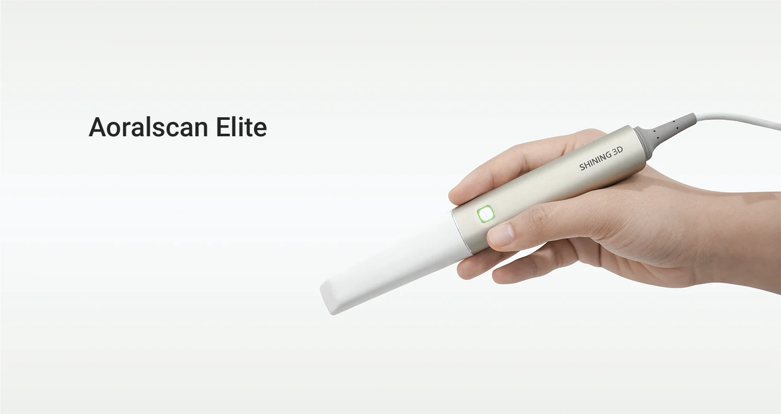
SHINING 3D DENTAL approaches this requirement by integrating intraoral photogrammetry with intraoral scanning in its Aoralscan Elite system. According to the company, the system can capture implant positions and angles using coded scan bodies while also collecting detailed intraoral and gingival data.
The two in one design is intended to simplify the acquisition process. Instead of using one system to capture implant positions and another to capture intraoral surfaces, the necessary data can be collected through an integrated workflow.
The system is designed for applications including edentulous implant cases and All on X treatment. SHINING 3D DENTAL also states that its coded scanbodies support compatibility with more than 100 implant manufacturers.
For practices and laboratories considering a photogrammetry dental scanner, compatibility is an important factor because the scanner needs to work with the implant systems used in daily cases.
What Should Professionals Consider Before Adopting Photogrammetry?
Accuracy should be an important consideration, but it should not be the only one. A practical evaluation should include the complete workflow.
First, consider how the scanner records implant positions and whether the system provides appropriate scanbody support for the implant connections being used.
Second, evaluate how photogrammetry data is combined with intraoral scan data. A workflow that requires excessive manual alignment or additional equipment may reduce some of the efficiency gained from digital acquisition.
Third, consider usability in actual clinical conditions. Full arch implant scanning can be time sensitive, so scanning speed, tracking stability, ergonomics, software integration, and data export options can all affect the overall experience.
Finally, evaluate whether the system can support both complex implant cases and routine scanning. A device that can function as an everyday intraoral scanner as well as a photogrammetry system may provide greater practical value.
From Implant Position Capture to a Connected Digital Workflow
The growing interest in photogrammetry reflects a broader development in digital implant dentistry. The objective is not simply to obtain another type of scan, but to improve how clinical information moves from the patient to treatment planning, CAD design, and final restoration.
Recent research has continued to compare intraoral photogrammetry with conventional intraoral scanning and extraoral photogrammetry for complete arch implant applications, demonstrating the importance of evaluating accuracy within the specific clinical workflow.
For practices handling a growing number of full arch implant cases, a photogrammetry 3D scanner can provide a dedicated method for capturing implant relationships while maintaining detailed intraoral data. With its integrated IPG approach, SHINING 3D DENTAL offers a workflow designed to bring these functions together in one system.
As full arch digital dentistry develops, the value of photogrammetry will increasingly depend not only on measurement accuracy, but also on how effectively the technology fits into everyday clinical and laboratory processes.
